The information gap
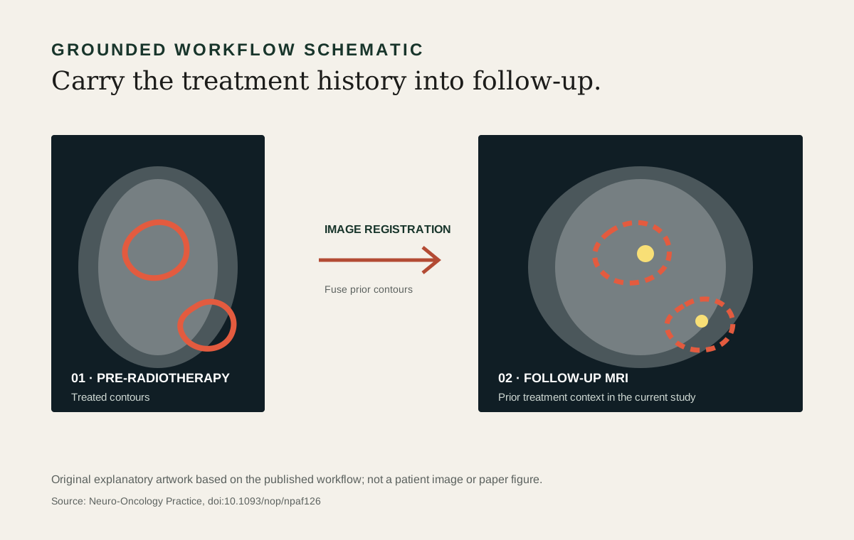
After radiotherapy, a follow-up magnetic resonance imaging (MRI) examination needs to be interpreted in light of what was previously treated. Treatment contours and current images can be separate pieces of the clinical record.
The co-authored framework brings pre-radiotherapy brain-metastasis contours into follow-up MRI to support that assessment.
Evaluation in a review workflow
The framework registered planning CT to follow-up MRI, propagated treated-lesion contours, and exported DICOM data compatible with a picture archiving and communication system (PACS). The study involved 40 patients, divided between geometric and physician-review studies, and evaluated review with the fused information. Average review time decreased from 7.97 to 3.95 minutes in the reported comparison.
The paper also examines agreement between physicians. Inter-physician agreement is a measure of consistency among readers, not a direct measurement of diagnostic accuracy against ground truth.
What the evidence supports
This was a retrospective single-centre evaluation. Anatomical change and image quality can affect registration. The result points to the value of presenting prior treatment information clearly during follow-up review. It is a workflow study, and the reported review-time change should be interpreted within that study’s setting and design.
The article was published online in December 2025 and appears in the June 2026 issue of Neuro-Oncology Practice.
Sources & further reading
Research summary updated October 5, 2026. Findings describe the linked study and do not establish clinical readiness beyond its evaluation.